Knee Diagnosis - The end ?
Comments
-
In a way both are right. The biggest use of MRI is almost certainly for soft tissue lesions such as tumours. But within a bone related specialty, MRI is of inestimable value for looking at subtle changes within bone which are not seen on plain X-rays.0
-
kayakerchris wrote:In a way both are right. The biggest use of MRI is almost certainly for soft tissue lesions such as tumours. But within a bone related specialty, MRI is of inestimable value for looking at subtle changes within bone which are not seen on plain X-rays.
Absolutely. The guy is saying bone isnt seen on MRI which is just false, if it was the case when I got a polytrauma into ITU I wouldnt get a report post MRI stating all the fractures seen. I believe most guidelines say that you should have an X-ray first and if this doesnt show anything and you are still sore an MRI should be performed. At the end of the day he's had MRI which in my mind and the mind of the 2 specialists I spoke to trumps getting plain X-rays.
Looking at his photos he's a radiographer and has probably spend too long looking at films and not enough time interpreting them.........Santa Cruz Blur XC
Nicolai Helius FR
Planet X Carbon RED0 -
oldwelshman wrote:If you want to discuss this in private I am happy to do so tehn you wont look so much of a prick.
In fact I know exaclty what I am talking about and if you want a lecture as to how mri images are acquired and recontructed and a lesson in mri physics I am happy to provide it to you. I do not need internet links to mri images I have thousands of themto look at. As you see tto like to browse then try looking at mri theory and you will see it is based on hydrogen protons though you can view others but thats not don often it is calles spectroscopy but I suppose you know that.
What you see in the images is not bone it is bone marrow and often you can see fractures and cartlidge damage to to the presence of fluids.
When rf is applied to a body at the lamor frequence it is the hydrogen protons filpped out of phase and bone does not have enough of these these which is why bone apopears black in mri as there is no signal given off as the protons relax back into line of magnetic filed, but I suppose with your superior knowledge you know this also.
What you see in the images is the bone marrow which has hydrogen protons, the bone appears very thin and black.
Maybe you can explain to me why you think bones are better scanned on mri as compared to x-ray.
One of the uses of mri for bones is for micro fractures and I thought you would have mentioned scaphoid injries.
These types of scans are in the minority and done mostly after other exams, the vast majority of mri images are for tumours, ortho, vascular, diffusion, muscular and some functional mri but I guess you also know this.
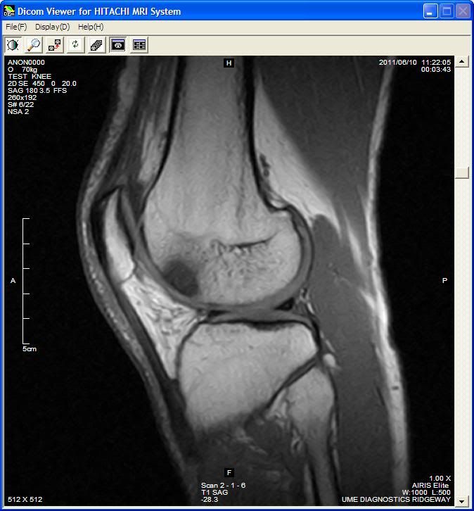
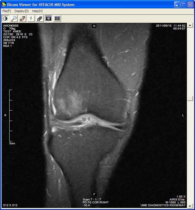
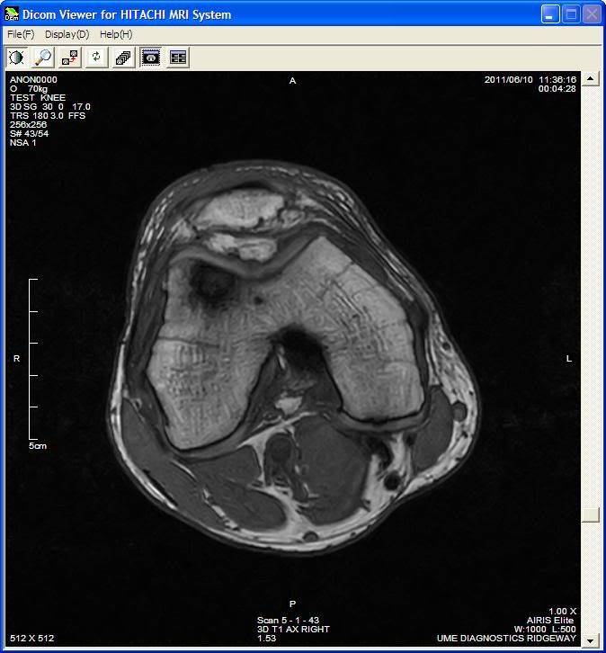
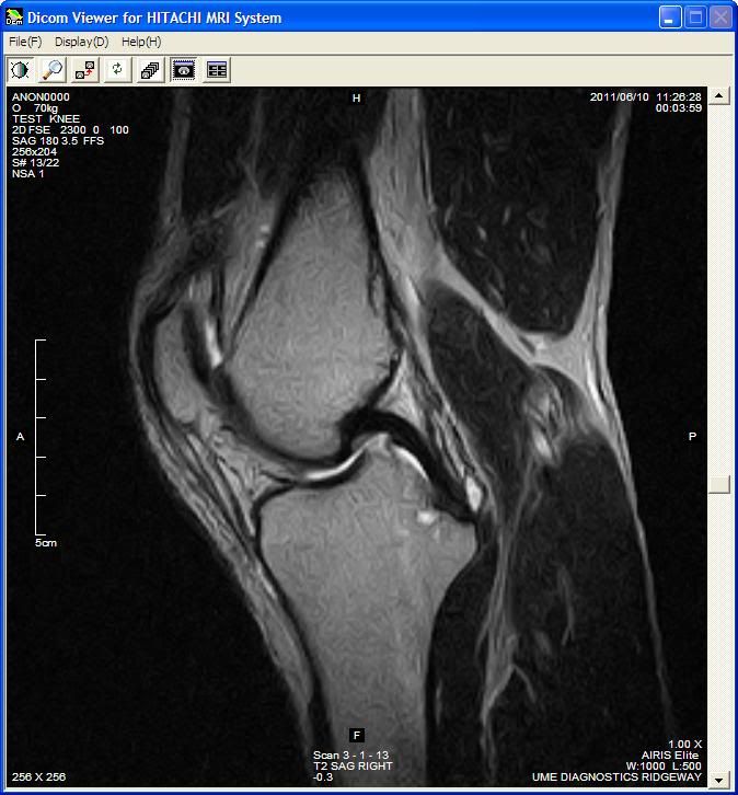
Maybe with your expert knowledge you can give me your diagnosis for my knee below and with your knowledge maybe you will identify what scan technique each one is, then I will be impressed :
:


You contradict yourself here. You say that bone cant be seen/is difficult to see but then you mention scaphoid. Given your extensive knowledge you will know that this is because the scaphoid carries a high risk of the complication of avascular necrosis. Scaphoid fractures are so commonly missed on plain films most countries opt for MRI to diagnose the fracture. In this country we opt to treat clinically on first presentation and x ray twice to confirm, not exactly gold standard.
Im sure that you also know that cervical fracture can not be excluded in patients with midline tenderness without a CT scan as once again plain x rays will not exclude a fracture
If a straightforward bony injury is suspected an x ray will always be useful and is a standard investigation. It can not exclude all types of fractures and will miss a great deal in the spectrum of bone disease. MRI is infinitely more sensitive particularly more helpful when there could be an overlap in the cause/diagnosis between bone and soft tissue.Santa Cruz Blur XC
Nicolai Helius FR
Planet X Carbon RED0 -
Last post
 it is not false that you cannot see bone in mri, bone itself does not give of signal it is absolutely black, the signal you see is ingress of fluids or bone marrow or other none bone tissue. This is what can indicate micro fractures.
Anyway my knee still has bone bruise and I will be having micro fracture surgery at some point, but think I will wait until after next years world masters track champs as I don't fancy six weeks sat up followed by longish rehab.0 -
hi folks - O/P here.....just an update for you.
Seen a physio - who spent the whole of the first session prodding, twisting, pulling, almost every muscle on my leg...he spent so much time ....he didn't have time to give me any specific excercises ! 2nd visit (with the mri result) he told me not to be too down beat - much could done to improve the function of my knee - by first loosening of the tightness that he felt was present all down my leg, hamstrings, calves, IT bands, deep glutes, pirifomis. - he gave me some stretches to do 3 times daily. I've been doing these for just over a week and the pain has subsided quite a lot. at times i think ive got it beat - then a get a twinge - which can be very disheartening.
taking calcium, glucosamine, rosehip supplements - and wear a knee support first thing in the morning - or if I know i am going to do a bit of walking.
done 2, 7 mile bike rides - without any reaction - although i get a twinge on any sort of moderate incline, and if i attempt any sort of out of the saddle.
got to go back in a couple of weeks - for some strengthening exercises and possibly taping the knee.
but at the moment I can get around pretty well - and I guess if I didn't have aspirations to get back on the bike in a meaningful way - i would consider it job done. But I guess the dilemma is do i risk cycling (something i enjoy) - v the risk of more permanent damage, loss of mobility, and permanent pain in later years0 -
My wife gets a twinge every now and then, doesn't stop me cycling.
Keep at it Kingrollo, you know you want to 0
0 -
I have long standing knee problems and find that cycling is one of the best things for me to do. I have had 6 operations (3 on each knee) and know that knee joint replacements will be needed, unfortunately for me is that I am not even 30 and my surgeon wont do a joint replacement until I am 60.
I have had my medial patello femoral ligaments reconstructed with a graft which is like having an elastic band holding the knee caps in place, this has been fine whilst cycling but it looks like I may have snapped my right graft twisting to get milk out of the fridge to make a brew. Got a long recovery.
I would say just take it easy and know your pain threshold. I really hope you can continuing cycling 0
0 -
Hi. I also had knee problems due to excessive weight squatting at the gym. Had a couple of arthroscopes but still the pain reared its ugly head. I stopped leg work at the gym and concentrated making my legs stronger on the bike. I have had no pain whatsoever whilst cycling. I use quite a high cadence and try not to mash the gears. so far so good. But I am no medical expert so i think it wise that you listen to the doctors. i would recommend a pro bike fit as it corrected my left knee that was not tracking in the correct position. Is your complaint degenerative?0

